Study shows virtual colonoscopy works just as well as traditional kind
New York ? Having an X-ray to look for signs of colon cancer may soon be an option for those who dread the traditional scope exam.
Two of the largest studies yet of “virtual colonoscopy” show the experimental technique works just as well at spotting potentially cancerous growths as the more invasive method. It’s also quicker and cheaper.
The X-rays can help sort out who really needs the full exam and removal of suspicious growths, called polyps. In one study, only 8 percent of patients had to have follow-up traditional colonoscopies, which are done under sedation and carry a small risk of puncturing the bowel.
But what some people consider the most unpleasant part can’t be avoided: drinking laxatives to purge the bowel so growths can be seen.
Still, proponents hope that the newer test will lure those who have balked at getting conventional screening.
“This is ready for prime time,” said Dr. Perry Pickhardt, one of the researchers at the University of Wisconsin Medical School who are reporting the results of their study in today’s New England Journal of Medicine.
A second, federally funded study at 15 sites around the country is meant to be the definitive test of virtual colonoscopy. Results have not been published, but they show the test to be promising.
Colonoscopies are recommended for everyone older than 50, but just about half get tested. Colon cancer is the nation’s second leading cause of cancer deaths, and an estimated 52,000 people will die from it this year. Screening can save lives by finding growths before they turn cancerous. Colonoscopies, considered the gold standard test, are recommended every 10 years and more frequently after polyps are found.
In traditional colonoscopy, performed by a gastroenterologist, a long, thin tube is inserted and snaked through the large intestines. Generally, any polyps that are spotted, regardless of size, are taken out in the process.
Virtual colonography uses a CT scanner to take a series of X-rays of the colon and a computer to create a 3-D view. A small tube is inserted in the rectum to inflate the colon so it can be more easily viewed. A radiologist then checks the images for suspicious polyps. Since the patient isn’t sedated, there’s no recovery time required.
But if any polyps need to be removed, the patient must then have a regular colonoscopy to do that.
National
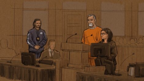
Man pleads guilty to killing a top Minnesota Democrat and her husband

New tornadoes outside Chicago as severe storms pummel the Midwest

Feds won’t seek death penalty in plea deal with man accused of killing top Minnesota Democrat

NASA unveils astronauts to test tech for future moon landing
Social Security’ fund faces shortfall one year earlier than expected
WASHINGTON (AP) — Social Security 's retirement trust fund is projected to face a funding shortfall in 2032, a ...