Ultrasound shows fetus in vivid 3-D
Dr. Paul Gisi and his wife, Shawna, haven’t had the pleasure of holding their baby boy yet.
But, with the help of some high-tech wizardry, they know exactly what he looks like.
“We can see his face. He’s so realistic — you can see his features so vividly,” said Shawna Gisi, 32, who is in the 37th week of pregnancy with her second child.
The couple are among the first Lawrence residents to take advantage of a new kind of service — Three-Dimensional Ultrasonography — offered to expectant mothers who are patients at Lawrence OB/GYN Specialists, 330 Ark.
Earlier this month, the four-physician practice, which is owned by Lawrence Memorial Hospital, began offering the most advanced technology available in 3-D ultrasound imaging, display and reproduction.
A new machine the practice has contracted to use can provide stunningly detailed and realistic pictures of babies who have reached at least the 23rd week of gestation.
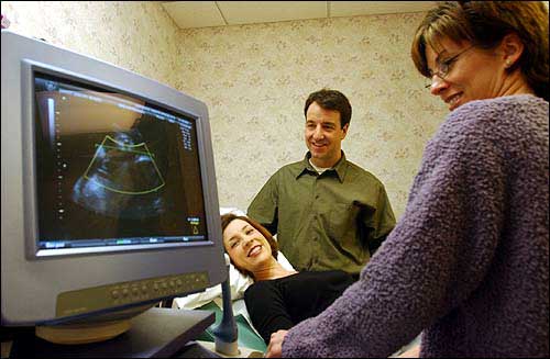
Paul and Shawna Gisi, Lawrence, look at an image of their baby boy with the help of sonographer Gayle Franklin. The technology behind Three-Dimensional Ultrasonography creates a more realistic image than ever for expectant mothers and their partners.
Since Jan. 3, about 10 of the clinic’s patients have undergone 3-D Ultrasound sessions, which last 20 to 30 minutes and are performed the same way as a traditional 2-D ultrasound.
Lawrence Family Practice Center, 4951 W. 18th St., also is able to provide 3-D ultrasound services. Pregnant women do not need to be patients of the center to schedule a 3-D Ultrasound session.
Shawna Gisi already has had two sessions of the 3-D ultrasound, one on the first day the clinic offered the technology, and another Thursday. Both times she was accompanied by her husband, Paul, 38, an obstetrician/gynecologist at Lawrence OB/GYN Specialists.
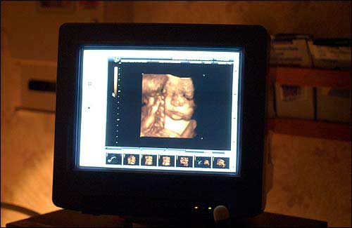
The Gisi baby, in his 37th week of gestation, is shown on the monitor via 3-D ultrasound.
While 2-D ultrasound remains the standard doctors rely upon for diagnostic and treatment purposes during pregnancy, 3-D ultrasound uses similar sound wave technology to offer expectant parents something extra: a clear look at their baby before it’s born.
“In our office, it’s going to be (used) for the ‘wow’ factor. Patients can take these pictures back to show family and friends that can be interpreted by non-medical people,” Paul Gisi said.

A 3-D ultrasound image captured Thursday clearly revealed the face of Paul and Shawna Gisi’s son, who doesn’t have a name yet. His eyes, nose, mouth and chin were perfectly visible.
“This is so exciting — I can see my baby!” Shawna Gisi said.